Typically, Microtomes Are Used To Slice Embedded Tissue into Very Thin Slices in Microbiology
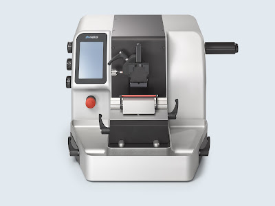 |
| Microtomes |
Microtomes are the most commonly used apparatus/instrument in
histology labs for obtaining sample sections for further microscopic
examination. Knife clamps, a sample holder, a thickness gauge, and other
accessories are included. It is a mechanical device that moves the sample
towards the blade/knife in small steps of a few microns in length to obtain the
required thickness section (thickness of the section mostly ranges from 5-10
nm). One of the most important tools in microtomy procedures is the knife
(microtome knife). In most cases, a wedge (C type) knife is used to cut the
sample section during a routine microtomy procedure.
The benefits provided by microtomes in histopathology practises
are expected to drive microtome growth. Microtomes are used in clinical and diagnostic
research laboratories, as well as industrial labs, to obtain routine paraffin
sections and plastic-embedded specimens, high-quality sections with less
fatigue, and to ensure the safety, reliability, and consistency of results.
This has created a significant opportunity for manufacturers to create
innovative microtomes using cutting-edge technologies.
According to Coherent Market Insights, The global Microtomes
Market is estimated to be valued at US$ 128.5 million in 2019 and is
expected to exhibit a CAGR of 6.2% during the forecast period (2020-2027).
A Microtome is a specialised
precision cutting instrument that slices sections from a block of embedded tissue
accurately and repeatedly. Different types of microtomes, as well as
specialised microtomes, are used to section paraffin and plastic-embedded
tissues and frozen tissues. A sharp knife and a tissue block are held in a
fixed relationship in any microtome. Each pass of the tissue through the knife
advances the tissue block by a predetermined amount—the section thickness.
The thickness of frozen sections is typically 8 to 15 m, wax sections
4-10 m, and plastic histological sections 0.5-3 m. Sections for electron
microscopy must be extremely thin, about 200 times thinner than wax sections.
Plastic sections used in transmission electron microscopy (TEM) are typically
cut in the 60-100 nm range.
Samples must be chemically fixed
with aldehydes, dehydrated in ethanol, clarified with xylene, and embedded in
paraffin before microtome sectioning. Most samples are commonly referred to as
Formalin Fixed Paraffin Embedded (FFPE) samples because they are fixed in a 10%
formalin solution (composed of approximately 4% buffered formaldehyde solution
stabilised with methanol). The use of the chemicals described will result in
overall poor ultrastructural preservation.
This technique, however,
preserves tissue microstructure and is still considered the gold standard for
pathology assessment in human and animal disease models. When planned sample
collection for CLEM is not possible, pre-prepared FFPE samples must be
investigated further. Although not ideal for ultrastructural preservation,
these samples can still be used for CLEM to answer specific biological
questions. When possible, it is strongly advised to use electron microscopy
grade aldehydes that have not been stabilised with methanol for sample
fixation.


Comments
Post a Comment